Cheng Lab
From model organisms to medicine: inside tissues in three dimensions.
This year we’re going cancer‑forward: starting with prostate virtual histology (label‑free PBCT micro‑CT), then ending with the “other organisms” that power the methods — octopus, zebrafish, and sentinel species.
In the room: ask to try the Sony ELF SR2 spatial display + hand controls (Neuroglancer 3D panel), or explore interactively on iPads.
From sample to screen: propagation‑based phase‑contrast micro‑CT
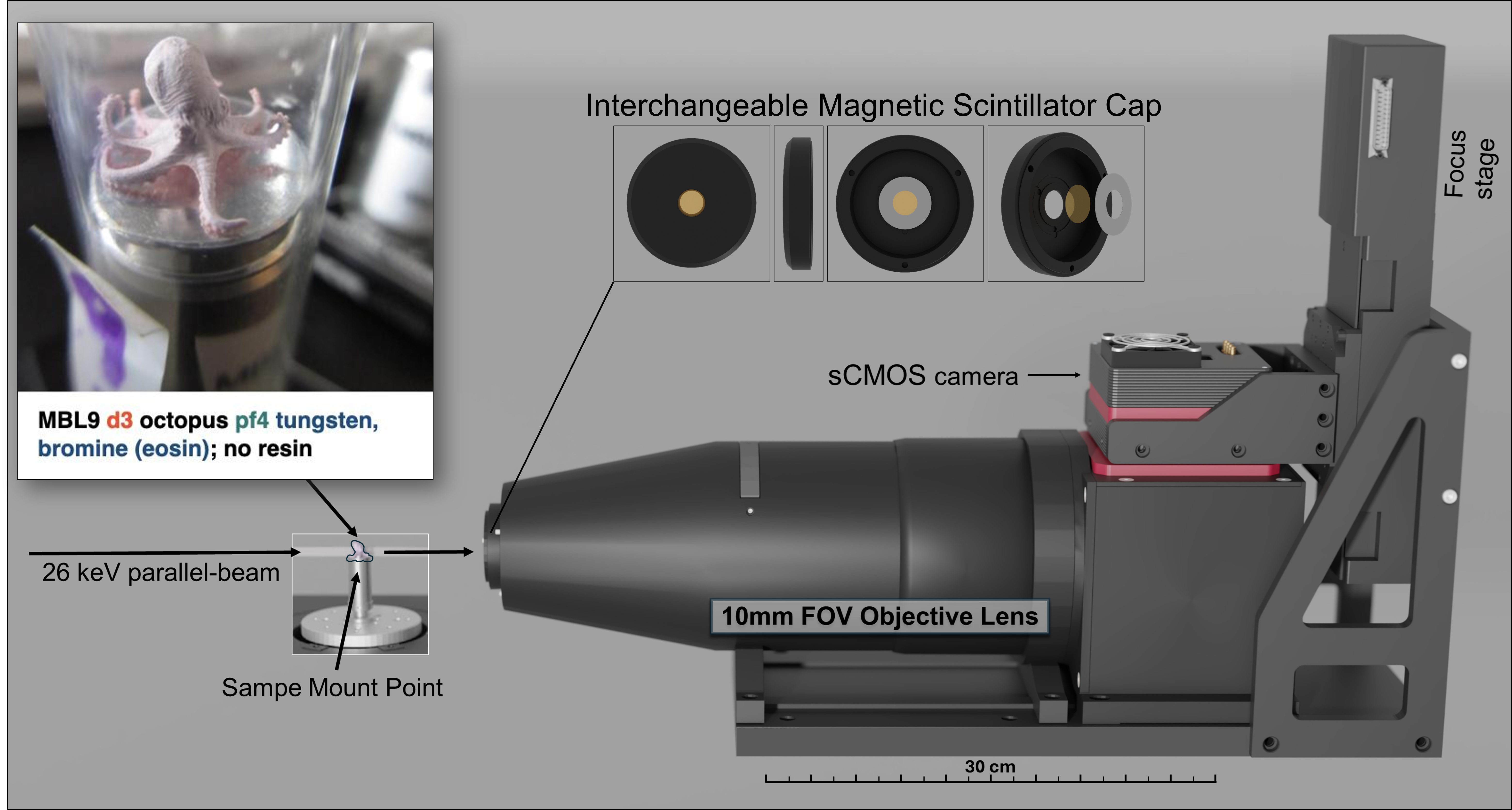
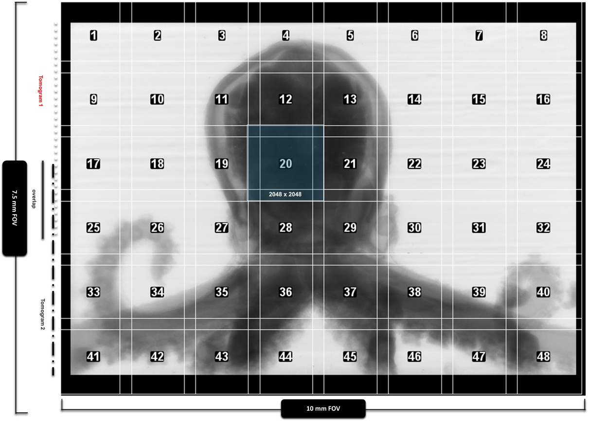
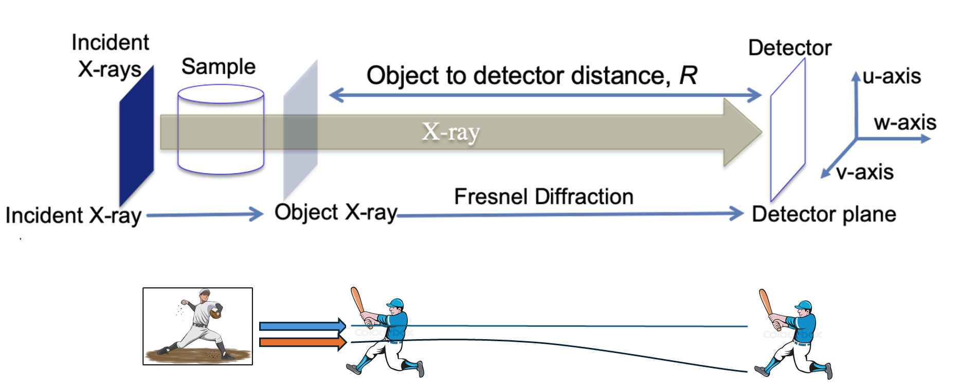
The goal: capture histology‑like detail across an intact specimen, then make it explorable in the browser—without physical sectioning.
Workflow (quick):
1) Intact sample (e.g., FFPE prostate core; stained model organisms when helpful).
2) Propagation‑based phase contrast (PBCT): edge enhancement from Fresnel diffraction at a chosen sample→detector distance.
3) Reconstruction + phase retrieval (single‑distance methods like BAC) to turn projections into a clean 3D volume.
4) Stitch / reslice: isotropic voxels mean you can cut any plane and generate 5 µm “virtual sections”.
5) Share + explore: Neuroglancer for interactive 2D/3D navigation on iPads or the Sony spatial display.
In short: one scan → many virtual sections → interactive 3D, with a pipeline that keeps the specimen intact for correlative H&E after imaging.
Label‑free 3D virtual histology of FFPE prostate biopsies
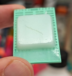
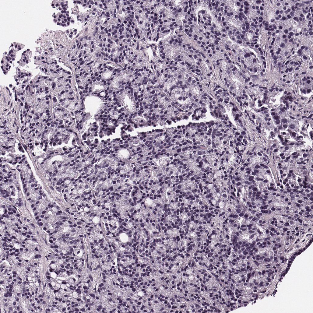
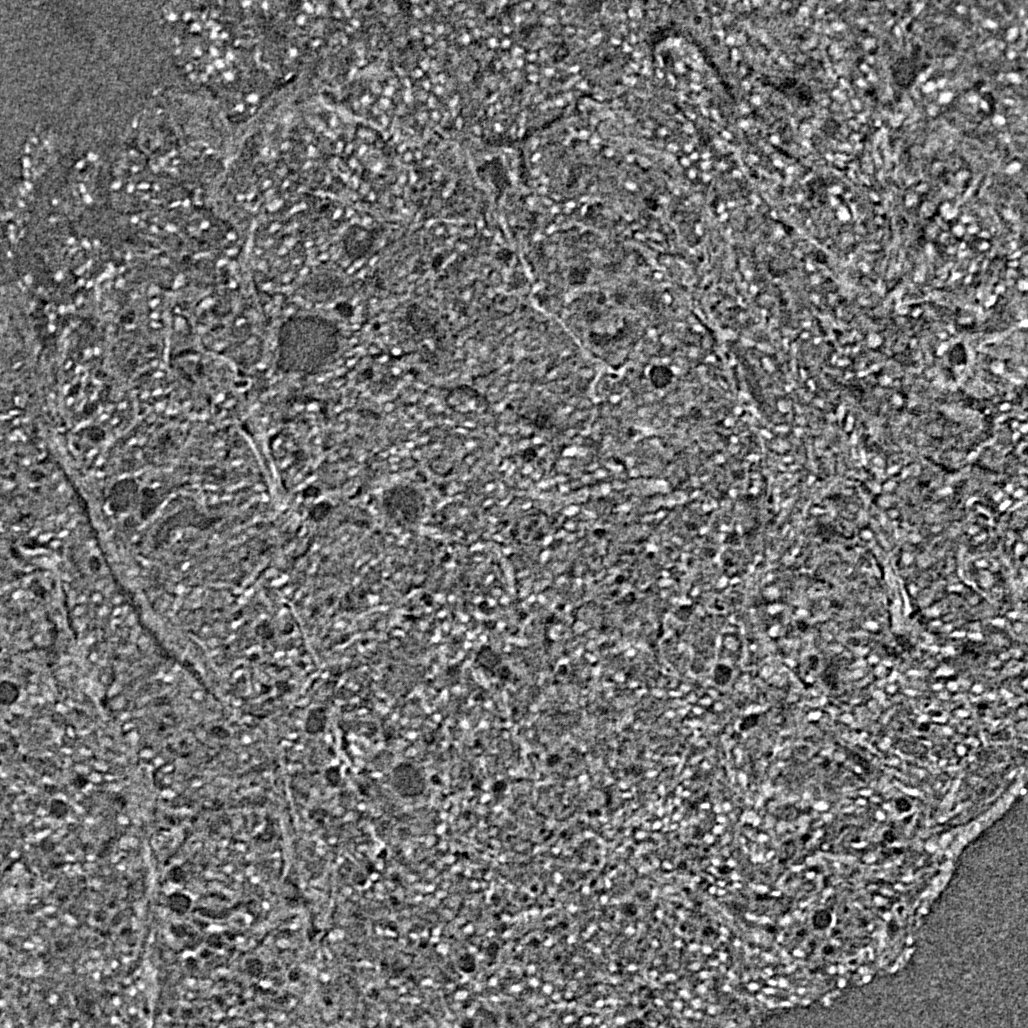
Tip: on iPad, tap the arrows to step through sample → H&E → PBCT → false‑color.
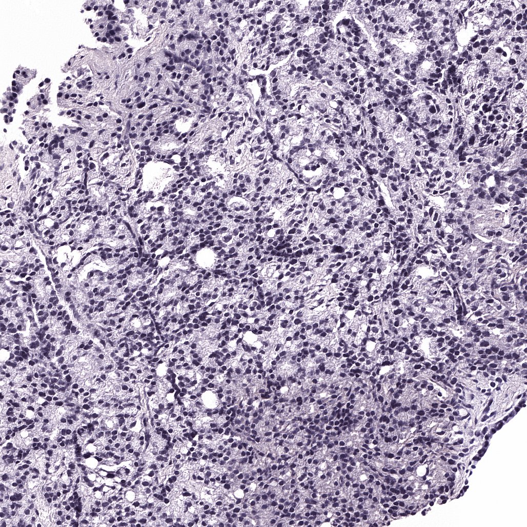
Why this matters: routine prostate grading is based on 2D sections that sample only a small fraction of a core. With propagation‑based phase‑contrast micro‑CT (PBCT), we can image the full biopsy volume nondestructively and explore it as a true 3D specimen.
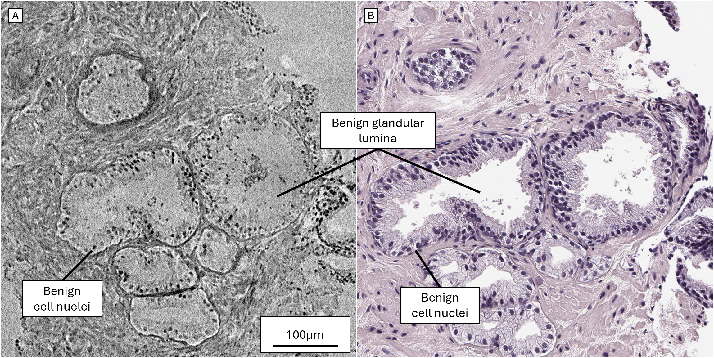
Our approach images residual paraffin‑embedded (FFPE) needle‑core biopsies without deparaffinization or added stain, producing isotropic, micron‑scale 3D data. We generate histology‑like virtual views (e.g., 5 µm maximum‑intensity projections) and correlate directly with serial H&E sections cut after micro‑CT.
From the draft manuscript: in these virtual sections we can distinguish benign gland patterns versus major diagnostic categories (Gleason patterns 3–5), and we share the full 3D volume through a customized Neuroglancer interface. Nuclear and gland segmentation/quantitation are also possible with sparse training data.
What to look for in 3D (fast tour):
• Gland continuity across depth (join/branch patterns that are hard to infer from one slice)
• Cribriform‑like architecture as a true 3D structure (not just a 2D snapshot)
• Spatial heterogeneity: how phenotype changes with depth and section angle
• Early computational overlays: nuclei/gland maps for rapid navigation
Note: cGAN false‑color is an active research prototype; we use it to test whether “pathologist‑friendly” color can speed pattern recognition while keeping the underlying 3D physics intact.
Whole‑core context that complements routine pathology
Care is where the research points: label‑free PBCT imaging of standard FFPE blocks can complement routine H&E by showing patterns across the entire core—without the undersampling and cutting artifacts inherent to physical sections.
We focus on integration: imaging residual paraffin blocks, correlating to existing histology, and sharing volumes in a secure, familiar web viewer.
Research use; de‑identified samples; no patient‑identifiable data are displayed.
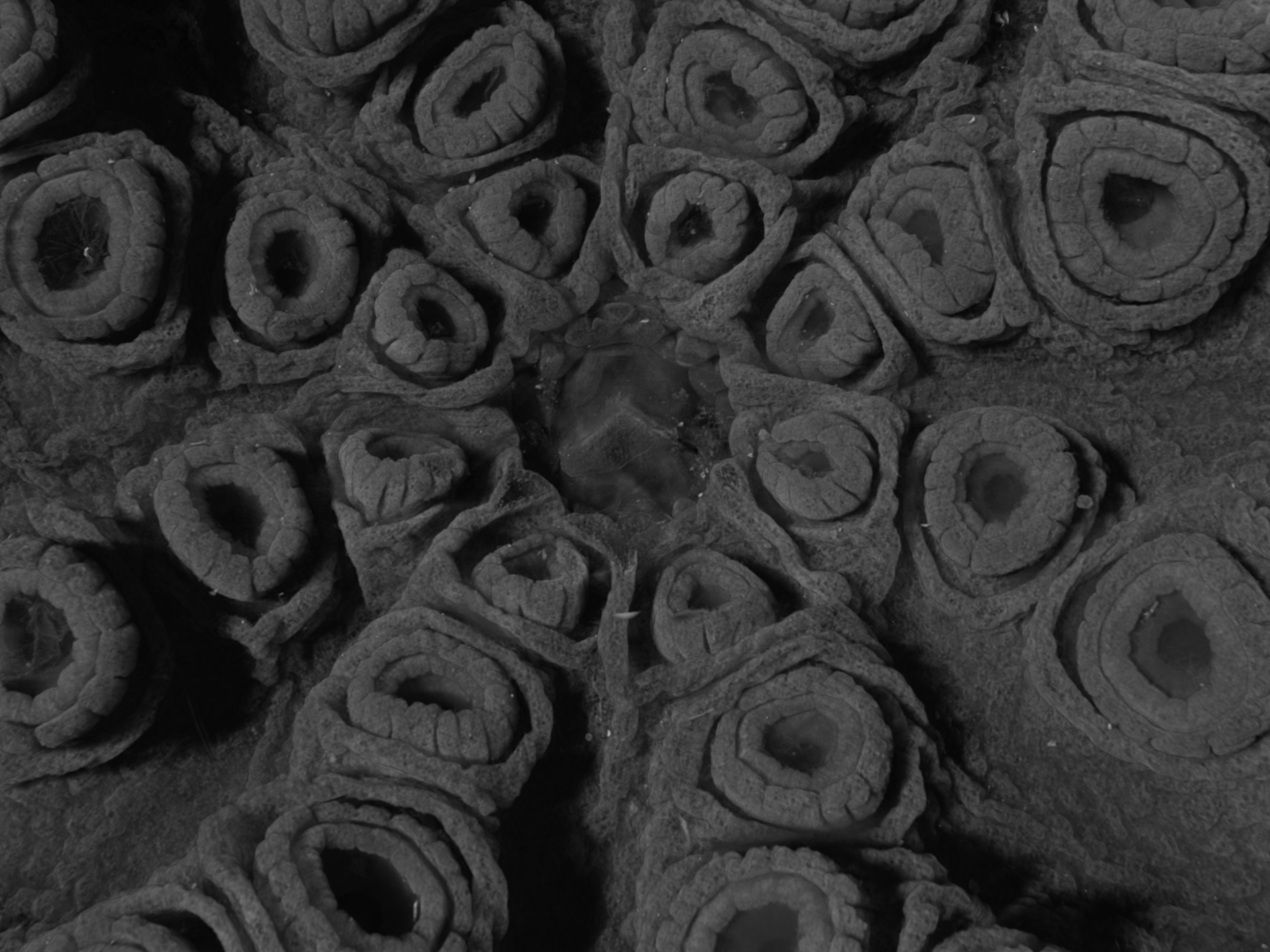
Octopus hatchling: whole‑body 3D anatomy you can navigate
Our metal‑stained octopus hatchling dataset turns an intact organism into an interactive 3D specimen. It’s a “methods‑meets‑biology” demo: long‑range pathways and organ relationships become intuitive when you can slice any way and render in 3D.
In-person: this is especially fun on the Sony ELF SR2 spatial display with hand controls.
Try this: follow a pathway from arm → brain, then switch to 3D rendering to see branching in context.

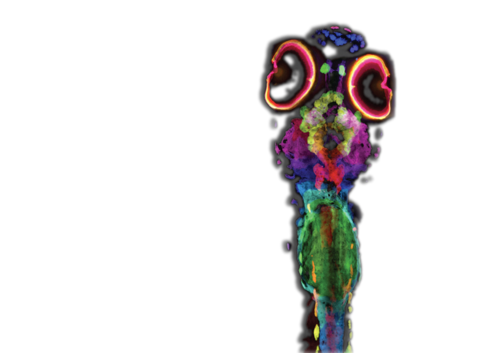
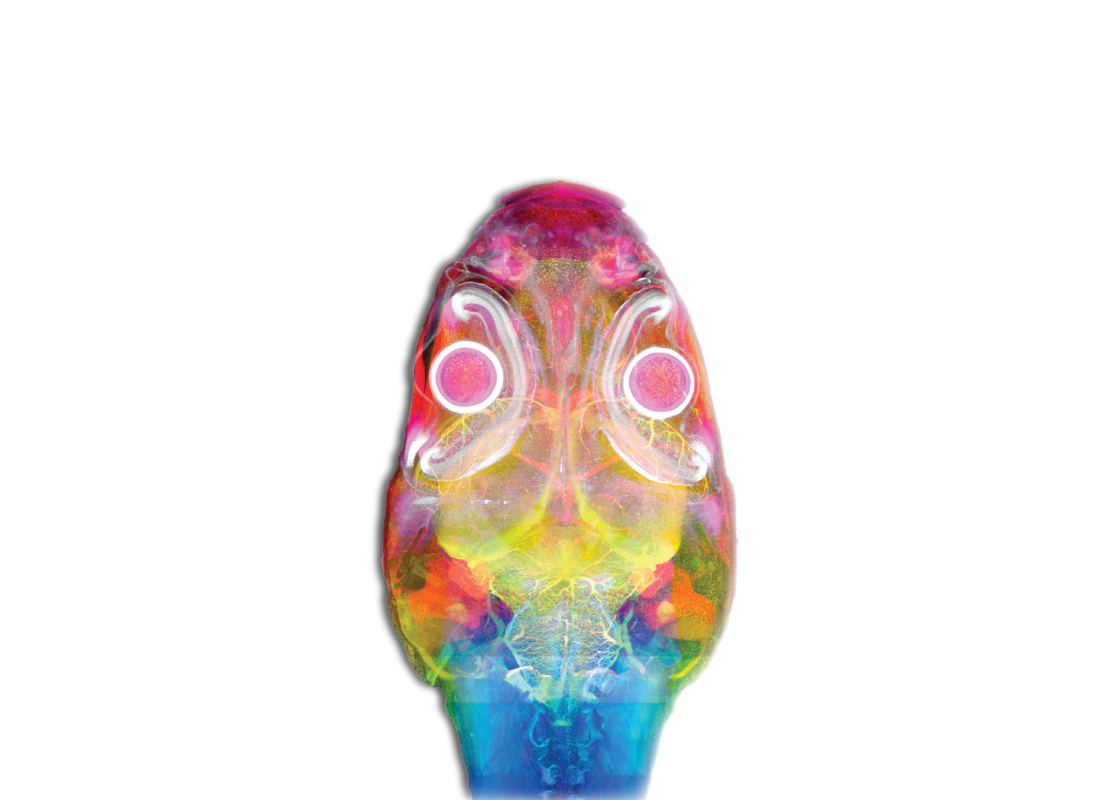
Zebrafish fly‑through training (skills that translate)
For education, we use whole‑organism datasets to teach spatial tissue intuition: learners can fly through retina, brain, and gut—then take those navigation skills into human tissue problems.
Bridge to cancer: the same browser‑based 3D viewing + annotation mindset used for zebrafish is what makes whole‑core prostate exploration feel natural (and fast).
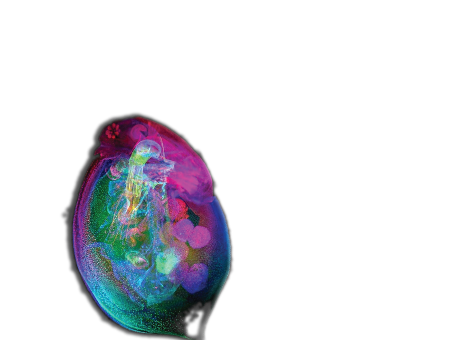

Sentinel species: prevention starts upstream
Prevention isn’t only screening—it’s understanding exposures and early tissue responses before disease. Sentinel species can reveal subtle, system‑level tissue changes that help define what “normal” looks like so that change stands out.
Cancer connection: environmental and lifestyle exposures can shape risk; atlases and quantitative microanatomy help link exposure science to prevention.
Questions? Want to collaborate?
Tell us what resonated — prostate/cancer, 3D methods, training, or prevention — and we’ll tailor a follow‑up.
Demo tip: append ?kiosk=1 to keep links in one tab and enlarge tap targets on iPad.