Wide-field acquisition
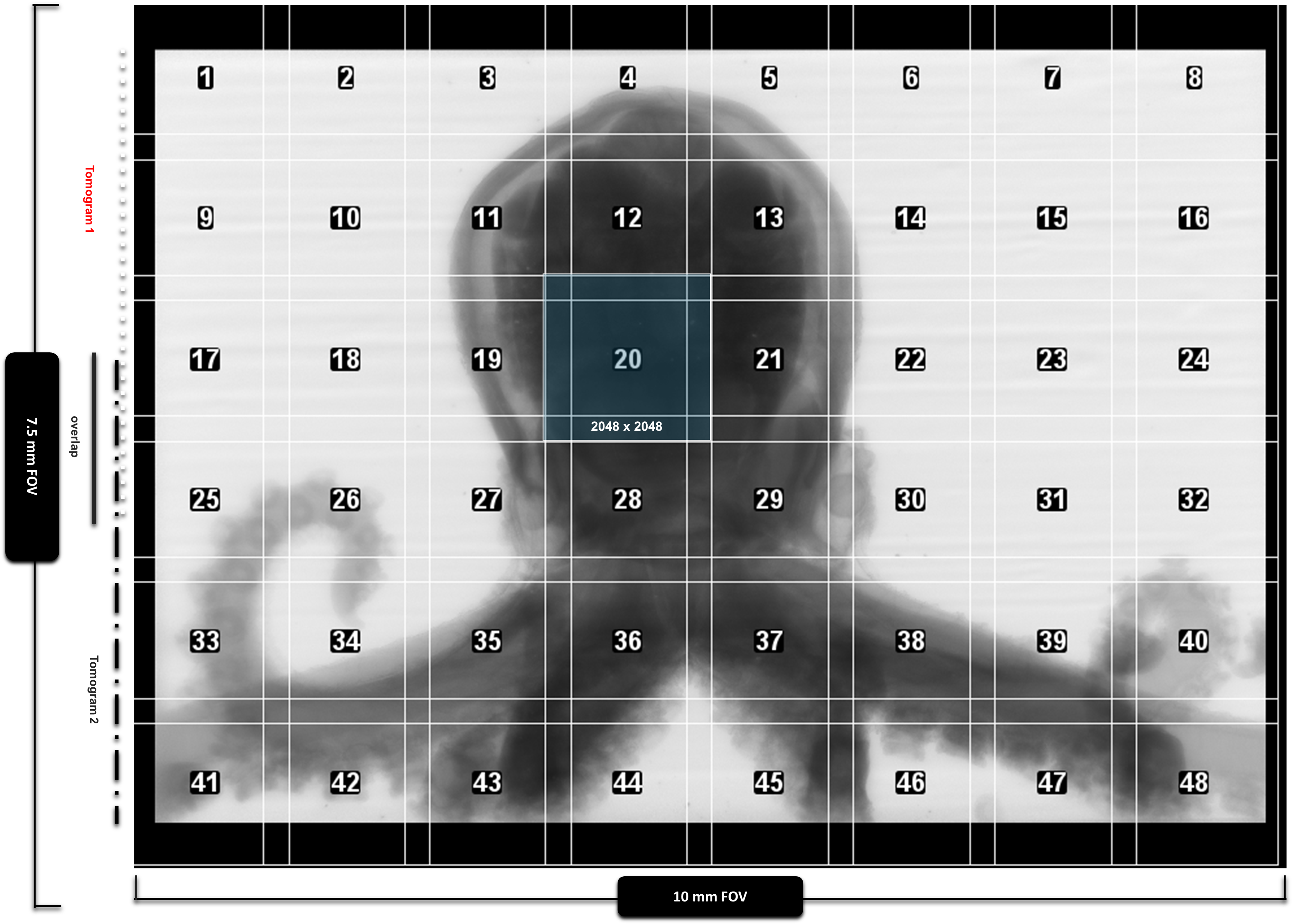
The detector captures a field large enough for the intact hatchling while retaining submicron sampling for microanatomy.

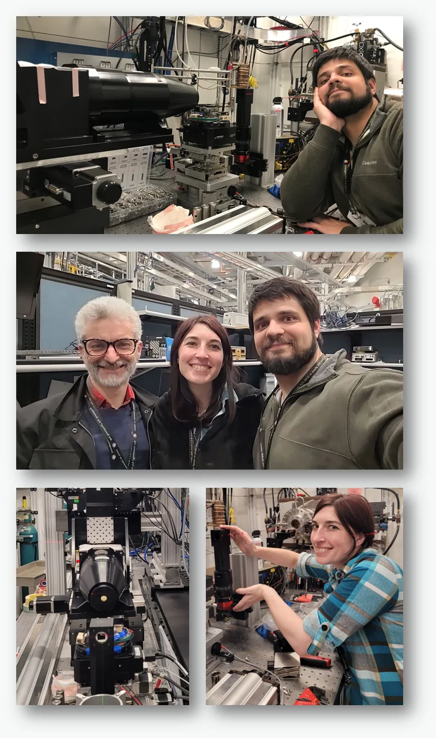
Custom wide-field detector at APS 2-BM
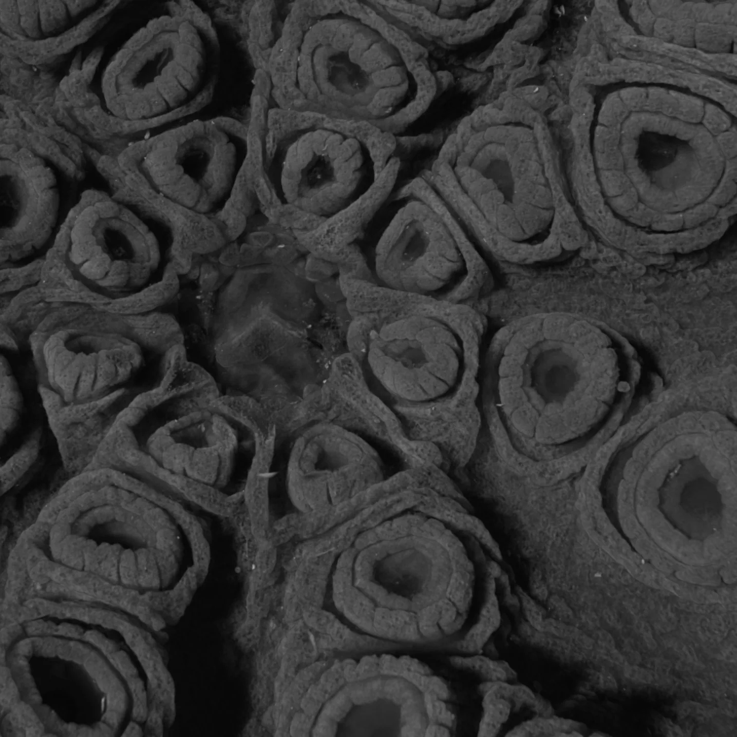
A 10 mm field of view at 0.7 µm isotropic voxels reveals intact neural architecture in a hatchling Octopus bimaculoides, from sucker-associated tracts to the arm-body transition zone.
The technical advance
Soft-tissue microCT usually trades field of view for resolution. The APS 2-BM wide-field detector extends histotomography into a centimeter-scale field while preserving histology-like contrast for intact biological specimens.
The result is a whole-organism volume that keeps long-range anatomy continuous. Instead of reconstructing a picture from disconnected fragments, viewers can move through the same intact specimen from arms and suckers to the nerve ring and central brain.
Detector to digital specimen
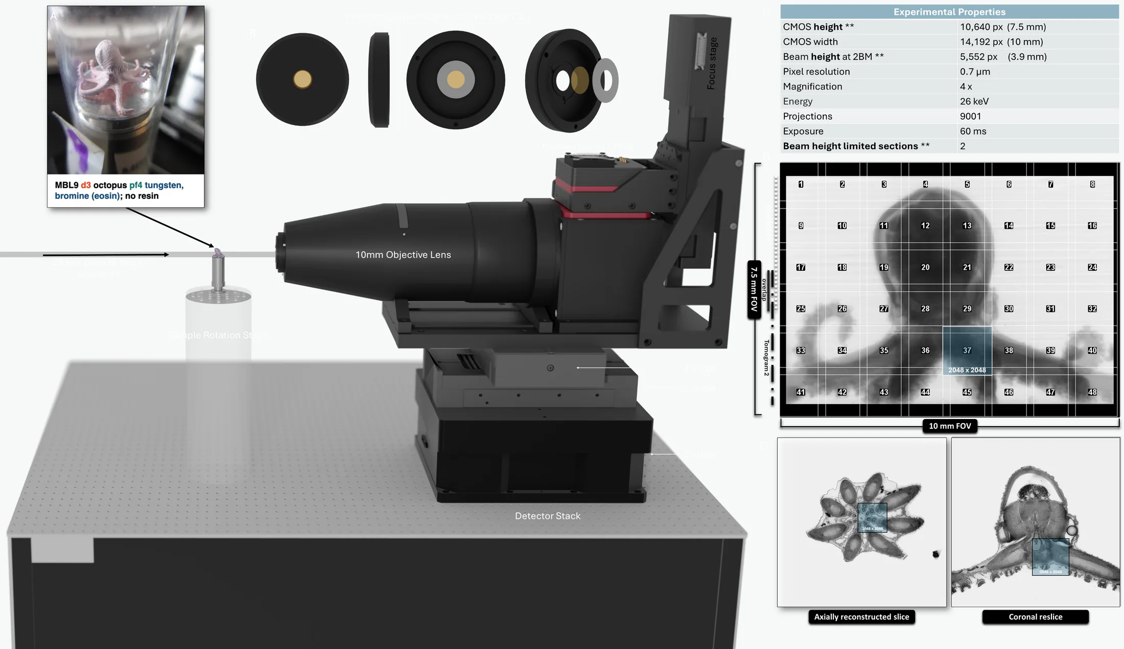
Acquisition, correction, reconstruction, segmentation, and web release are treated as one continuous pipeline, because each step affects whether long-range relationships remain interpretable.
The detector captures a field large enough for the intact hatchling while retaining submicron sampling for microanatomy.
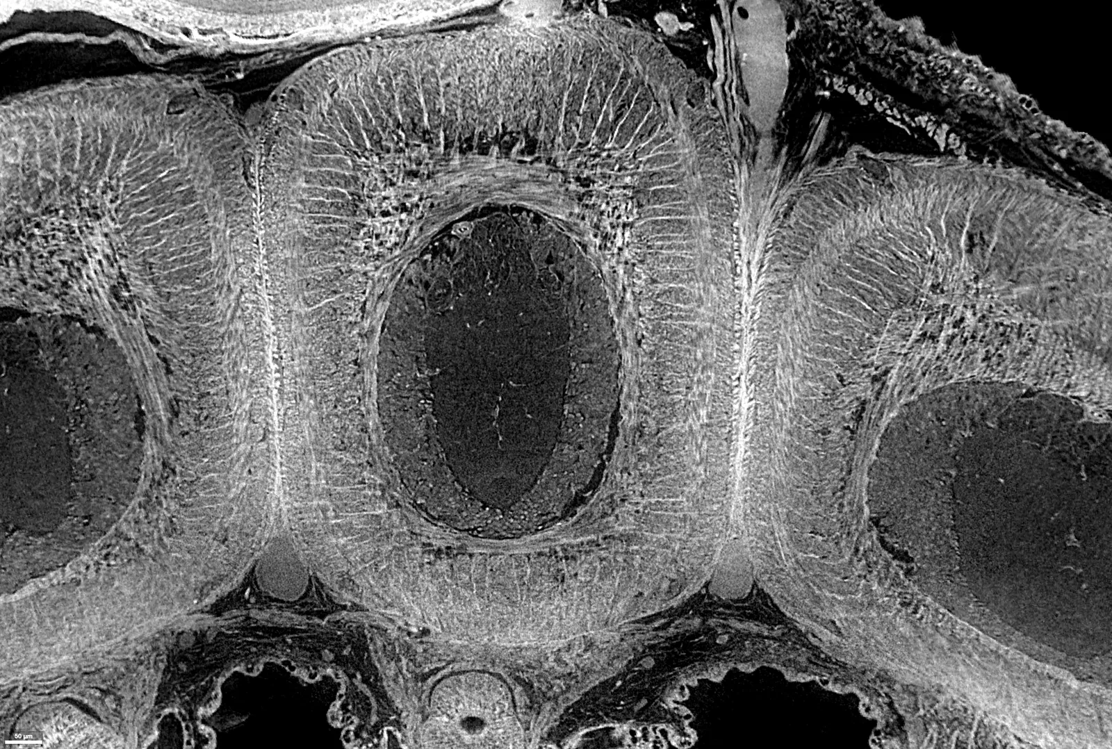
Sample drift, stripes, and ring-like artifacts are corrected so tissue boundaries and tract continuity remain readable.
The multi-terabyte specimen is released through browser-based viewers so attendees and collaborators can inspect the live volume.
Biological payoff
The octopus is a demanding test case because its arm nervous system, inter-arm commissures, and brain-bound tracts only make sense when they can be followed through the intact arm-body junction.
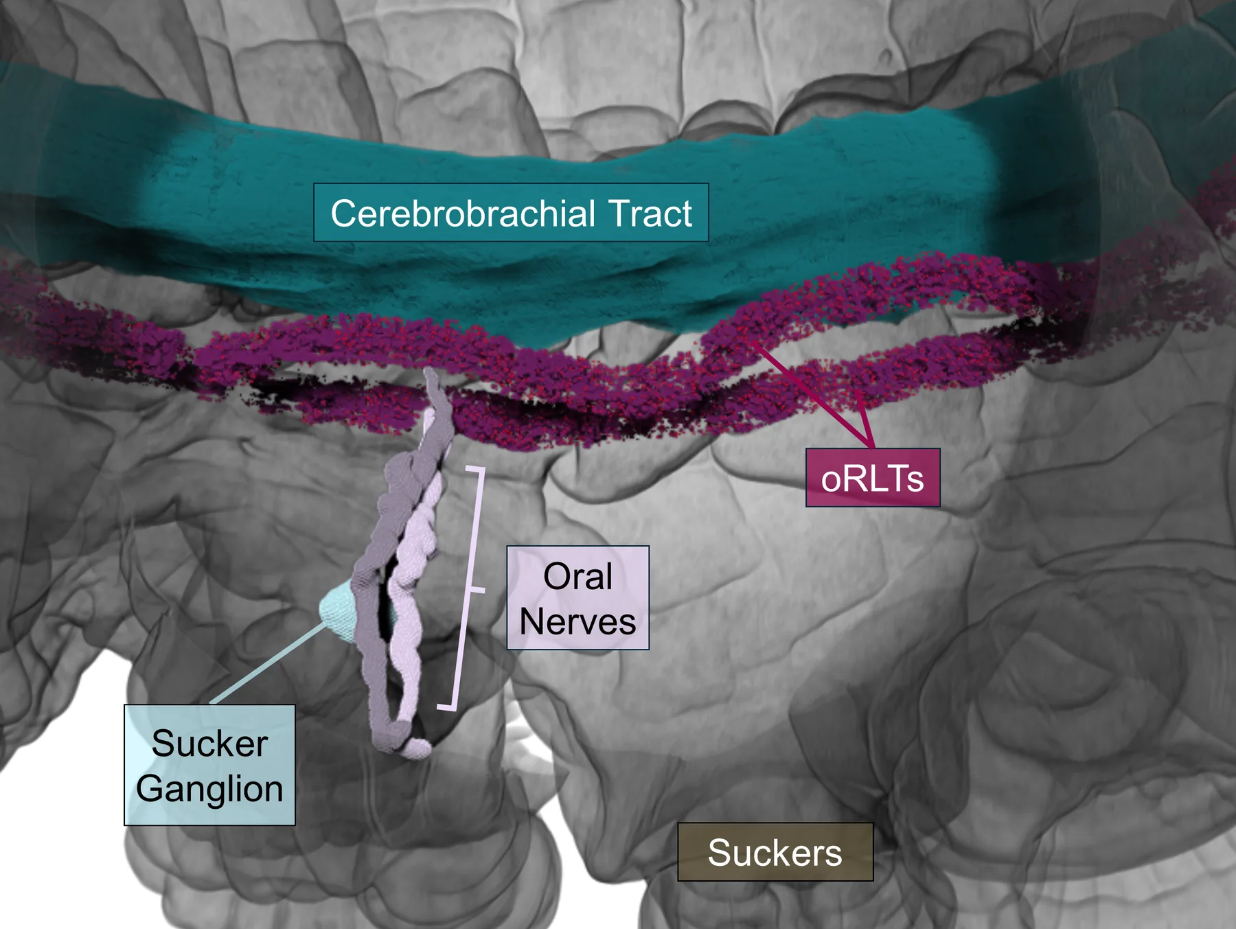
Longitudinal renderings show paired oral-root-associated longitudinal tracts within the axial nerve cord neuropil. They run parallel to the cerebrobrachial tract, repeatedly associate with oral nerve roots at successive suckers, and remain spatially separate over much of the arm.
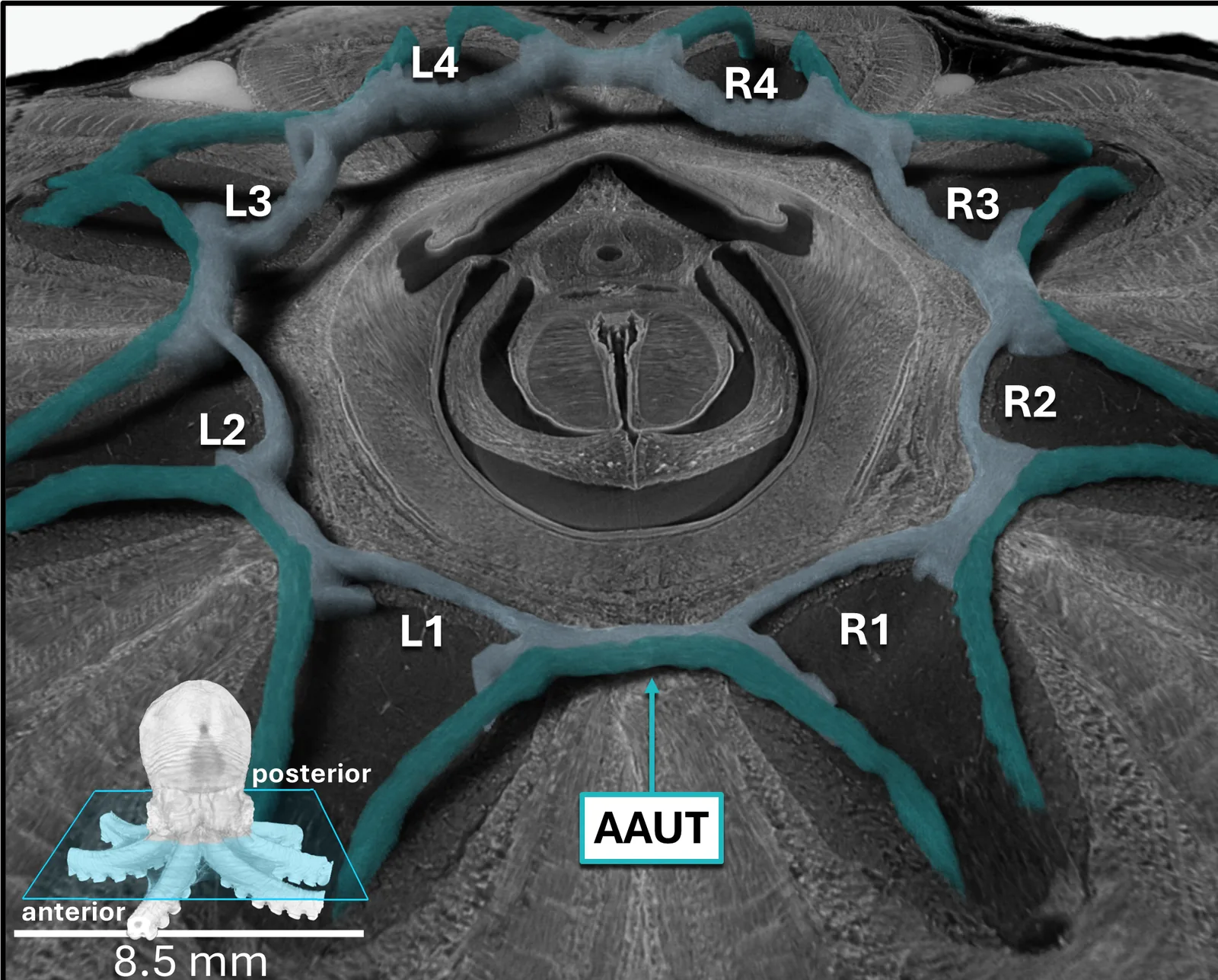
Within the nerve ring, arm-to-arm U-tracts form a repeated motif around adjacent arm bases. These fascicles follow a U-shaped route from one arm toward the next, rather than jumping between distant arms.
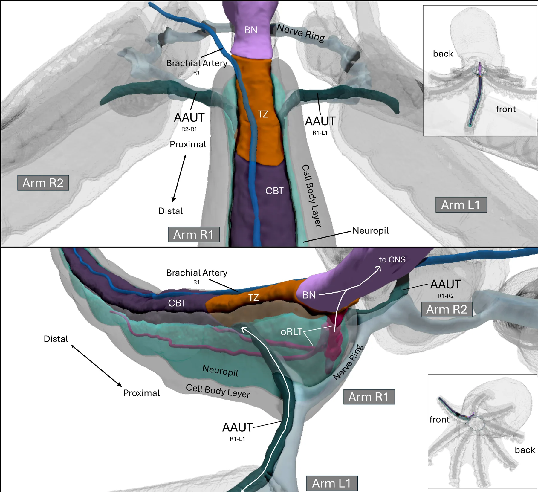
At the proximal arm-body junction, the brachial nerve and CBT form a transition zone. oRLTs approach from the axial nerve cord neuropil and enter the oral-medial brachial nerve, while AAUT fascicles enter laterally and continue into lateral CBT components.
Poster abstract
Submicron soft-tissue microCT usually trades field of view for resolution, limiting intact-organism imaging. We developed and deployed a custom wide-field detector at APS 2-BM that extends soft-tissue microCT to a 10 mm field of view at 0.7 µm isotropic voxels.
Using two scans and artifact-corrected reconstruction, we generated a whole-organism volume of an intact hatchling Octopus bimaculoides with histology-like soft-tissue contrast and segmented more than 300 structures across organ systems.
The octopus provides a demanding biological test because intact whole-animal continuity reveals long-range relationships that are lost in fragmented preparations, including paired sucker-associated longitudinal tracts in the axial nerve cord, repeated neighboring-arm fascicles within the nerve ring, and a proximal brachial nerve/cerebrobrachial tract transition zone.
The multi-terabyte dataset is being released as an open, interactive digital specimen, illustrating how APS-enabled wide-field microCT can connect detector development, beamline acquisition, reconstruction, and biological discovery.
For poster attendees
Open the Neuroglancer viewer to inspect the volume directly, or start from the Octo9 landing page for the broader data release. The most useful first move is to follow a nerve bundle from an arm into the proximal transition zone.